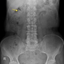
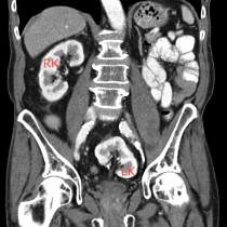
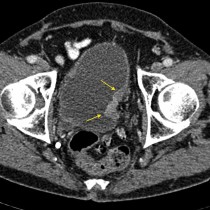
• Close
    • Home
    • Videos
    • Tutorials
    • Gallery
    • Links
    • FAQs
    • All Case Studies
    • Diagnostic Imaging
      • Imaging Modalities
        • Radiography
        • Fluoroscopy
        • Ultrasound
        • CT
        • MRI
        • Nuclear Medicine
      • Contrast Media
      • Radiological Anatomy
      • Case Studies
      • PACS
      • Radiation Safety
      • Radiological Descriptive Terms
    • Interventional Radiology
      • Introduction to IR
      • Image-guided Biopsy
      • Case Studies
    • UCD Students
      • Weekly Rotation
      • Radiology Conferences
      • Resources
      • Undergraduate Medals
    • Electives
    • Careers
    • About Us
St. Vincent's University Hospital Radiology Department
  • Diagnostic Imaging
    • Imaging Modalities
      • Radiography
      • Fluoroscopy
      • Ultrasound
      • CT
      • MRI
      • Nuclear Medicine
    • Contrast Media
    • Radiological Anatomy
    • Case Studies
    • PACS
    • Radiation Safety
    • Radiological Descriptive Terms
  • Interventional Radiology
    • Introduction to IR
    • Image-guided Biopsy
    • Case Studies
  • UCD Students
    • Weekly Rotation
    • Radiology Conferences
    • Resources
    • Undergraduate Medals
  • Electives
  • Careers
  • About Us
  • Home
  • Videos
  • Tutorials
  • Gallery
  • Links
  • FAQs
  • All Case Studies

Staghorn calculus

Staghorn calculus

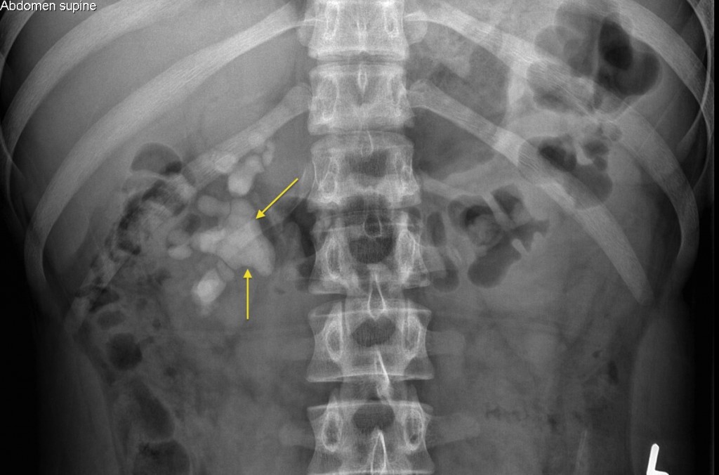
Staghorn calculus. This radiograph shows a staghorn calculus filling the renal pelvis and calyces of the right kidney in this 16 year old boy. These calculi are usually composed predominantly of struvite (magnesium ammonium phosphate), mixed with calcium phosphate. They are typically encountered in patients with recurrent urinary tract infections.


Related Case Studies

  • Renal calculus
  • Emphysematous cystitis
  • Ureteric calculus on CT KUB
  • Gallstone – ultrasound
  • Pelvic kidney
  • TCC bladder
Back
  • Interventional Radiology
  • Diagnostic Imaging
    • Emergencies
    • Neuro
    • Tubes & Lines
    • Trauma
    • Oncology
    • Hepatobiliary
    • Genitourinary
      • Horseshoe kidney with RCC
      • Xanthogranulomatous pyelonephritis (XGP)
      • Prostate cancer – MRI
      • Ovarian dermoid cyst
      • Epididymo-orchitis – ultrasound
      • Polycystic ovarian syndrome
      • Drug paraphernalia in vagina
      • Pelvic kidney
      • TCC bladder
      • Testicular tumour
      • Renal cell carcinoma
      • Polycystic Kidney Disease (2)
      • Polycystic Kidney Disease
      • Ovarian dermoid
      • Emphysematous cystitis
      • Obstructing bladder TCC
      • Retrograde urethrogram
      • Vesicoureteric reflux
      • Transitional Cell Carcinoma
      • Ureteric calculus on CT KUB
      • Staghorn calculus
      • Renal calculus
    • Gastrointestinal
    • Chest
    • Musculoskeletal
    • Vascular

All Case Studies

  • Main Categories

    • Interventional Radiology
      • Vascular IR
      • Non-Vascular IR
      • IR Oncology
      • Musculoskeletal IR
    • Diagnostic Imaging
      • Emergencies
      • Neuro
      • Tubes & Lines
      • Trauma
      • Oncology
      • Hepatobiliary
      • Genitourinary
      • Gastrointestinal
      • Chest
      • Musculoskeletal
      • Vascular
  • CONTACT US

    Department of Radiology
    St Vincent’s University Hospital
    Elm Park
    Dublin 4
    e.heffernan@st-vincents.ie

    Links

    • FAQs
    • Links

    SOCIAL

    © Copyright St Vincent's University Hospital Radiology Department 2025
    Web Design Dublin

    Search St. Vincent's University Hospital Radiology Department

    This website uses cookies. Accept Read More