Ovarian dermoid cyst
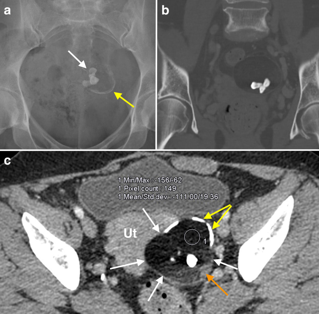
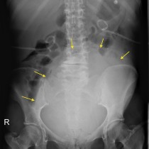
The peculiar abnormality on this 58 year old woman’s imaging was an incidental, asymptomatic finding. The radiograph (a), performed for a different reason, shows abnormal calcifications in the pelvis: there is some thin, curvilinear calcification indicated by the yellow arrow and a cluster of more chunky calcifications indicated by the white arrows. The latter actually represent teeth (molars in this case) and this tells us that the abnormality is an ovarian dermoid cyst (also known as a mature cystic teratoma).
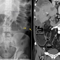
The coronal CT image (b), on bone windows, shows the three teeth more convincingly.
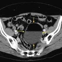
Image (c) is a transverse image from the same CT, on soft tissue windows. This shows the dermoid more clearly, the margins of which are indicated by the white arrows. It lies in the left adnexa, adjacent to the uterus (‘Ut’). You can see the curvilinear calcification in the wall of the mass that we spotted on the earlier radiograph (yellow arrows). This image also nicely demonstrates another typical feature of dermoids – while there is a small nodule of soft tissue density (orange arrow) at the edge of the dermoid, the rest of the mass is composed of fat. A circular region of interest (‘ROI) has been placed on the hypodense component of the tumour and the text above tells us that the mean density of this component is minus 111 Hounsfield units, and is therefore fat (review Hounsfield units here).
Ovarian dermoid cysts are common ovarian neoplasms which contain elements from multiple germ cell layers. These tumours are slow-growing and are usually an incidental finding in asymptomatic patients, most often of reproductive age. When large, they can result in ovarian torsion in which case the patient will present with acute pain. Malignant transformation can occur but is rare, therefore asymptomatic dermoids are usually managed conservatively unless they are large.
More than 90% of these tumours will contain fat which allows us to make a specific diagnosis on CT or MRI (and, in many cases, ultrasound). Around one third will contain teeth, and many will have other types of tissue such as skin or thyroid. Ovarian dermoids will be bilateral in around 10% of patients.
Reference: Saba et al. Mature and immature ovarian teratomas: CT, US and MR imaging characteristics. Eur J Radiol 2009;72:454-63. doi 10.1016/j.ejrad.2008.07.044.