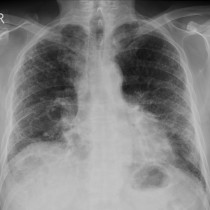
Scleroderma associated interstitial lung disease
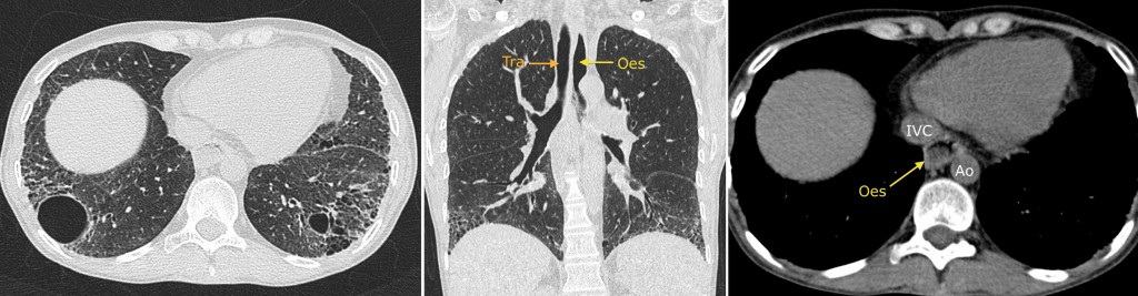
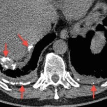
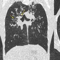
These transverse and coronal CT images, on lung windows, show basal-predominant interstitial lung disease with honeycombing and some larger cysts. You will also notice that on the coronal image and the right-hand soft-tissue window image, the oesophagus (‘oes’) is dilated, which hints at the underlying diagnosis here – scleroderma. If the oesophageal dilatation were not present, the differential diagnosis would be UIP, other connective tissue disease such as rheumatoid, asbestosis and drug-induced fibrosis (which can be caused by amiodarone, nitrofurantoin, methotrexate, bleomycin and many others). Pulmonary involvement is common in scleroderma although it is often asymptomatic. The lungs can also be involved secondarily if the patient is experiencing recurrent aspiration because of the oesophageal dilatation.
Ao = aorta; tra = trachea