Cystic fibrosis
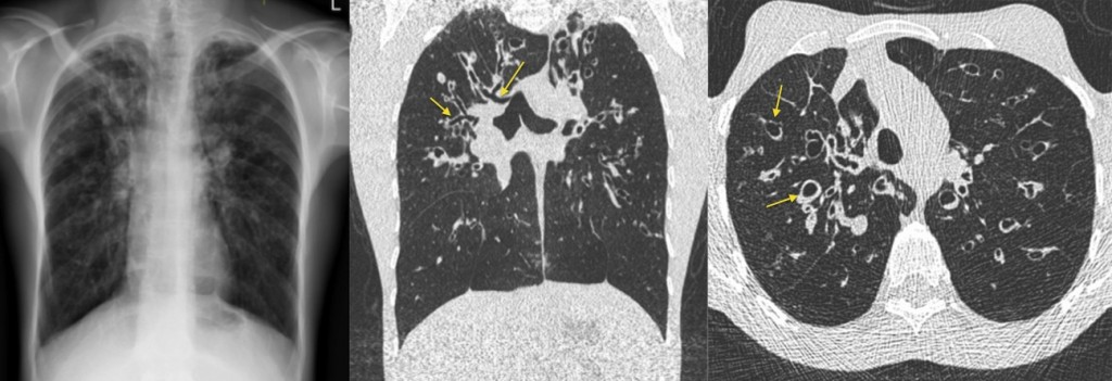
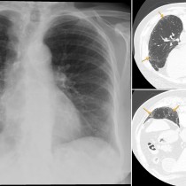
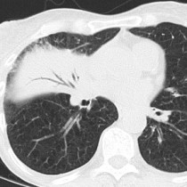
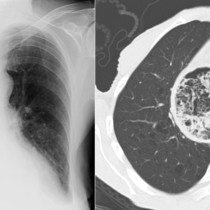
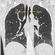
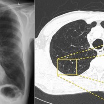
Cystic fibrosis. The CXR on the left shows the typical appearance of CF: hyperinflated lungs with increased markings in both upper zones, due to bronchiectasis and bronchial wall thickening. High-resolution CT is used for routine assessment of these patients, to evaluate for progression of the disease. In this example, the middle coronal CT image mirrors the CXR – the arrows indicate some of the dilated bronchi. The transverse CT image on the right shows how dilated these bronchi can become. CT will also show us areas of mucus plugging, active consolidation, mycetoma formation and small pneumothoraces (which can be loculated and very difficult to identify on a radiograph).