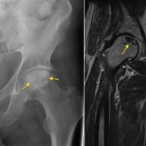
Interstitial lung disease – UIP
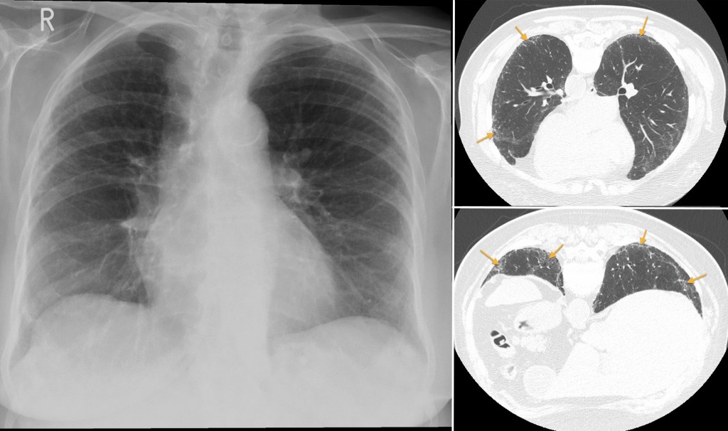
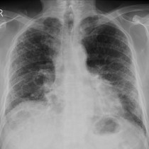
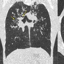
Usual interstitial pneumonia (UIP). This 78-year-old woman presented with progressive dyspnoea. The chest radiograph on the left shows how difficult it can be to identify early interstitial lung disease – it looks normal however the images from the patient’s CT (performed in the prone position) show subpleural, basal-predominant reticular opacities. In more advanced stages of UIP, CT often shows honeycombing, traction bronchiectasis (where bronchi become dilated because they are being pulled into areas of fibrosis), and volume loss.