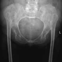
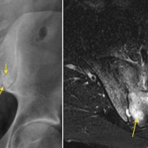
AVN – x-ray and MRI
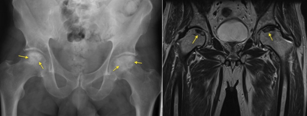
AVN. The plain film on the left shows typical appearances of osteonecrosis (avascular necrosis, AVN), with increased density of the femoral heads (arrows). The next stage in AVN is for the femoral heads to begin to collapse, following which secondary osteoarthritis will develop. Unfortunately, in the early stages of AVN, radiographs are usually normal. MRI is the most sensitive imaging test at all stages of AVN and should be requested whenever it is clinically suspected. The image on the right is from an MRI from the same patient, and shows low signal abnormalities in the superior aspects of both femoral heads.