Sacroiliitis – MRI
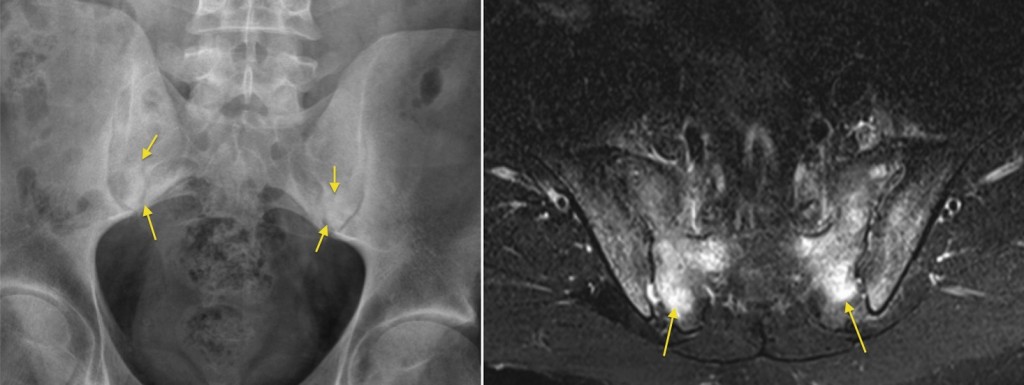
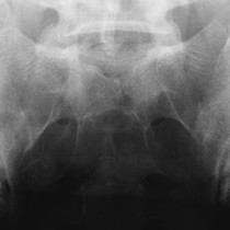
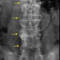
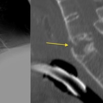
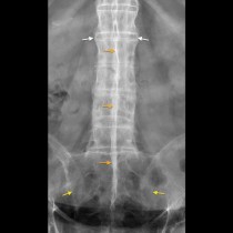
Sacroiliitis. The image on the left shows fairly subtle sclerosis (arrows) around both sacro-iliac joints, which could easily be overlooked, however the same patient’s MRI on the right shows extensive marrow oedema (the high signal indicated by the arrows), indicating that there is florid sacroiliitis (the patient had ankylosing spondylitis). MRI is far more sensitive than radiographs for inflammatory change at the SI joints and is routinely requested when sacroiliitis is suspected clinically but the patient’s plain films are normal.