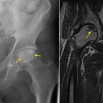
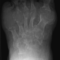
Osteomyelitis in diabetic foot
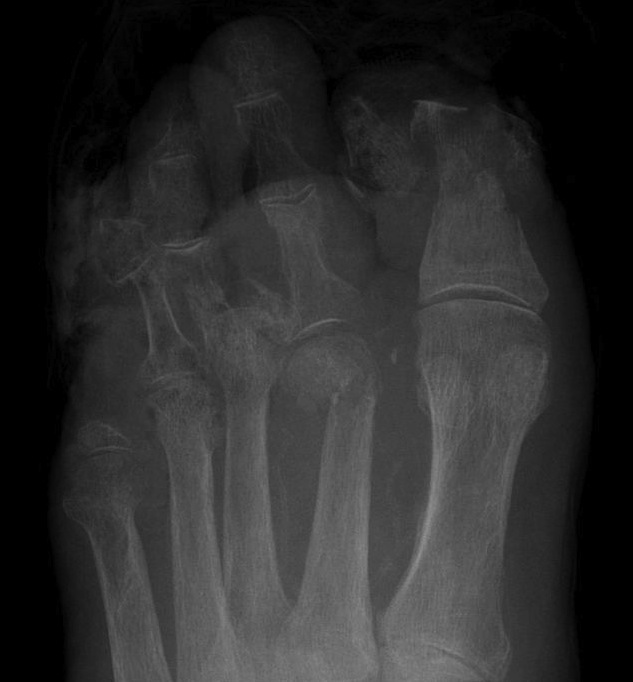
Osteomyelitis. This diabetic patient has had previous episodes of osteomyelitis – note previous amputation of his fifth toe. He represented with a large ulcer over his great toe. This radiograph shows complete destruction of the first distal phalanx and some destruction of the head of the proximal phalanx, indicating osteomyelitis. Further amputation was required. In the early stages of osteomyelitis, radiographs are usually normal – MRI with gadolinium is performed in such cases where osteomyelitis is clinically suspected.