Pulmonary fibrosis
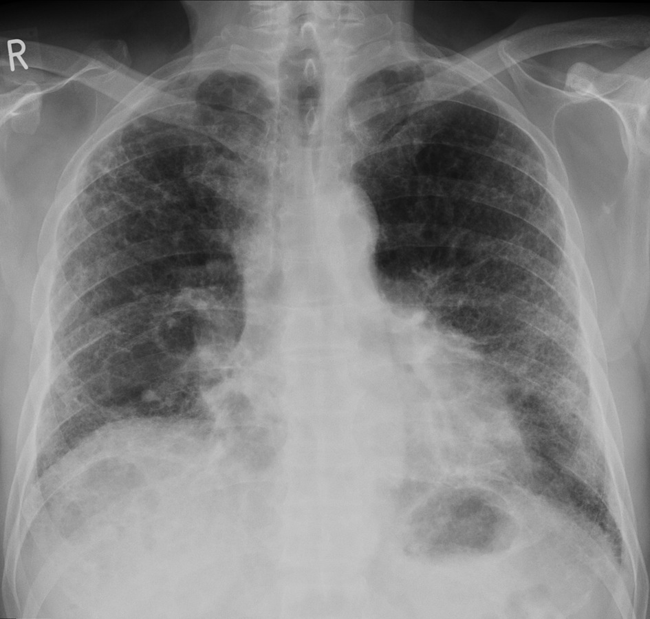
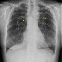
Pulmonary fibrosis. This CXR shows a ‘reticular’ (net-like) pattern of increased interstitial lung markings in the peripheries of both lungs, worst at the lung bases. There is also some evidence of volume loss – the right fifth anterior rib bisects the diaphragm (usually the sixth or seventh does this). HRCT would better assess the severity of the fibrosis. The differential diagnosis for this distribution of fibrosis would include IPF, fibrosis related to connective tissue diseases such as RA and scleroderma, drug-induced fibrosis (e.g. methotrexate), and asbestos-related fibrosis. This patient had IPF.