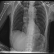
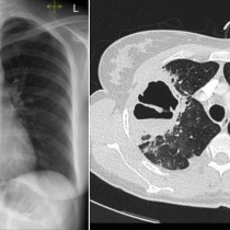
Aspiration pneumonia
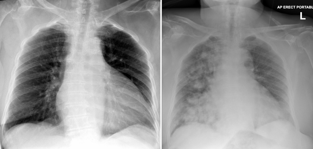
These are chest radiographs of an 83 year old female stroke patient, taken one day apart. On the image on the left the lungs are clear, but the next day the patient suddenly deteriorated and became hypoxic. The portable CXR on the right shows new dense consolidation in the right lung. The rapidity of the change and the distribution of the abnormality are clues to the underlying diagnosis – aspiration of gastric contents. This occurs most commonly in the right lung. When it occurs in the supine position, aspiration pneumonia tends to involve the posterior segment of the upper lobe and the apical segment of the lower lobe (both are involved in this example), while if the patient is erect when aspiration occurs it will favour the right middle and lower lobes.