Right middle lobe pneumonia
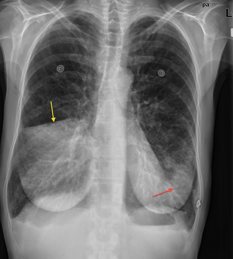
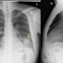
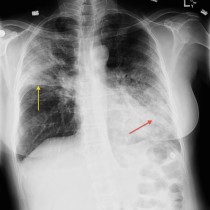
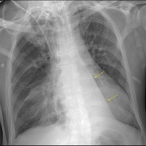
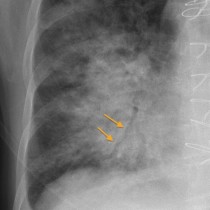
Right middle lobe pneumonia. This patient has consolidation in the right middle lobe. Note the increased density of the consolidated lung. The right hemidiaphragm remains visible, however the right heart border has disappeared, indicating a middle lobe process. Also helpful is the fact that the consolidation stops abruptly at the horizontal fissure (yellow arrow). There is also some consolidation in the lingula – we can say this because the increased density in the left lung is obscuring most of the left heart border (red arrow), but spares the silhouette of the left hemidiaphragm.