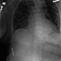
Malpositioned nasogastric tube
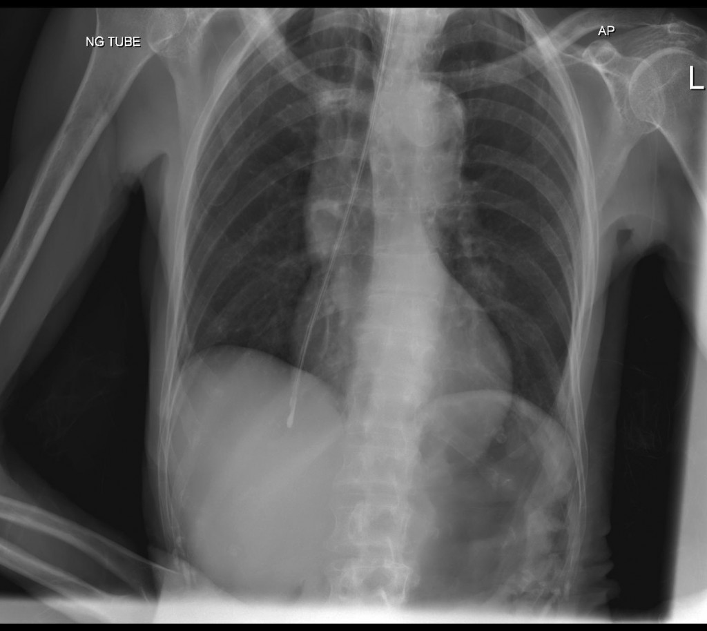
The NG in this patient has been inadvertently advanced down the right main bronchus into the periphery of the right lower lobe. This would obviously be unsafe to use for feeding, but also places the patient at risk of pneumothorax.