Epididymo-orchitis – ultrasound
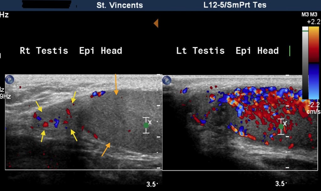
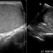
A common indication for scrotal ultrasound is for the diagnosis of epididymitis or epididymo-orchitis. At ultrasound, epididymitis typically manifests as a thickened, hypoechoic epipidymis that demonstrates increased blood flow when Doppler is used – occasionally, this hypervascularity is the only finding. In the example shown here, the image on the left shows the normal right epididymis (yellow arrows) and testis (orange arrows). In contrast, the left epididymis and testis show dramatically abnormal hypervascularity on Doppler. About 20-40% of patients with epididymitis will have evidence of orchitis on ultrasound. We will sometimes see a reactive hydrocoele in association with these findings. Clinically, the differential diagnosis in this setting is often testicular torsion – in established torsion, blood flow will be absent rather than increased.