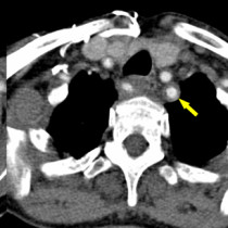
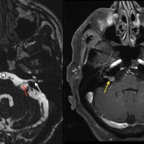
Carotid Stenosis
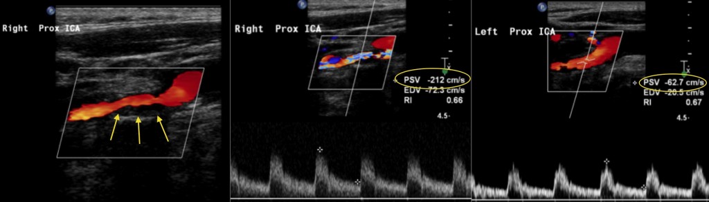
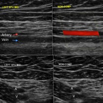
Carotid stenosis. These images are from a carotid Doppler study in a patient who was admitted following a stroke. On the colour Doppler image on the left, you can see narrowing of the lumen of the right internal carotid artery due to atheromatous plaque (arrows). The middle image shows a measurement of blood flow velocity through this stenosis, showing an elevated ‘Peak Systolic Velocity’, PSV, of 212 cm/sec. When above 125 cm/sec, we consider carotid flow velocity to be elevated. The higher the velocity, the greater the degree of stenosis. An image from the normal left internal carotid is shown on the right, where the PSV is only 62 cm/sec.