Polycystic ovarian syndrome
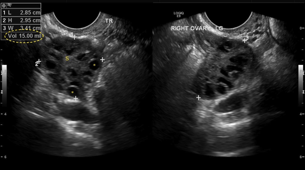
This 23 year woman presented with amenorrhoea and an elevated serum testosterone level. Her GP referred her for a pelvic ultrasound. These images are from that study and show transverse (left) and longitudinal (right) scans of her right ovary. This shows the classic sonographic appearances of polycystic ovarian syndrome (PCOS): the ovary is enlarged (normal is less than 10 ml, this one measures 15 ml in volume (circled on top left)), and there are multiple small anechoic follicles (some are marked with an ‘*’) in the periphery of the ovary – this is often called the ‘string of pearls’ sign. There is some hyperechoic stroma (‘s’) in the centre of the ovary. These findings are usually, but not always, bilateral. An abnormal ultrasound alone is not sufficient to make a diagnosis of PCOS – an additional clinical feature such as oligo- or amenorrhoea and/or supportive biochemical abnormality is also required.