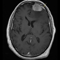
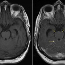
Acoustic neuroma
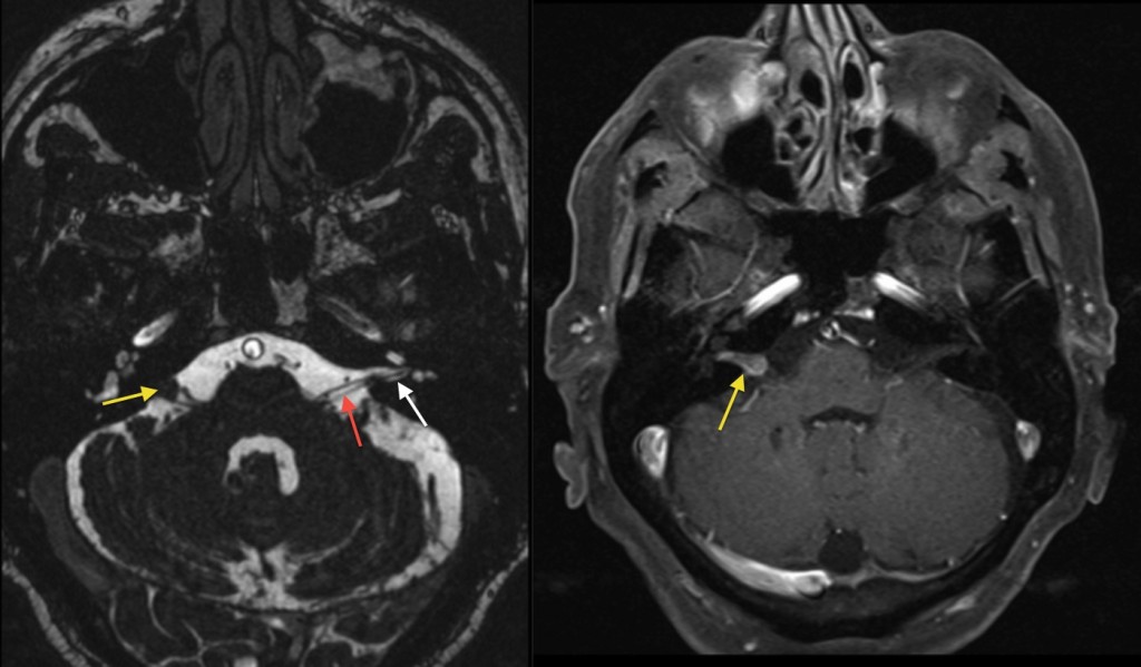
Acoustic neuroma. These images are taken from an MRI of the internal auditory meati (IAMs) in a patient with right-sided sensorineural hearing loss. The image on the left is a thin-slice T2-weighted MR which shows the normal internal auditory canal (white arrow), containing the 7th and 8th cranial nerves (red arrow). However, on the right side the canal is filled by a soft tissue mass which is protruding through the meatus into the cerebellopontine angle (yellow arrow). On the right is a post-contrast T1-weighted fat-saturated image showing uniform enhancement of the mass, which has the typical ice-cream cone shape of an acoustic neuroma (vestibular schwannoma). When bilateral, these tumours are diagnostic of neurofibromatosis type 2.