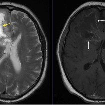
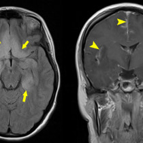
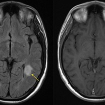
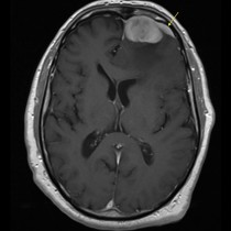
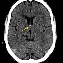
Meningeal metastases
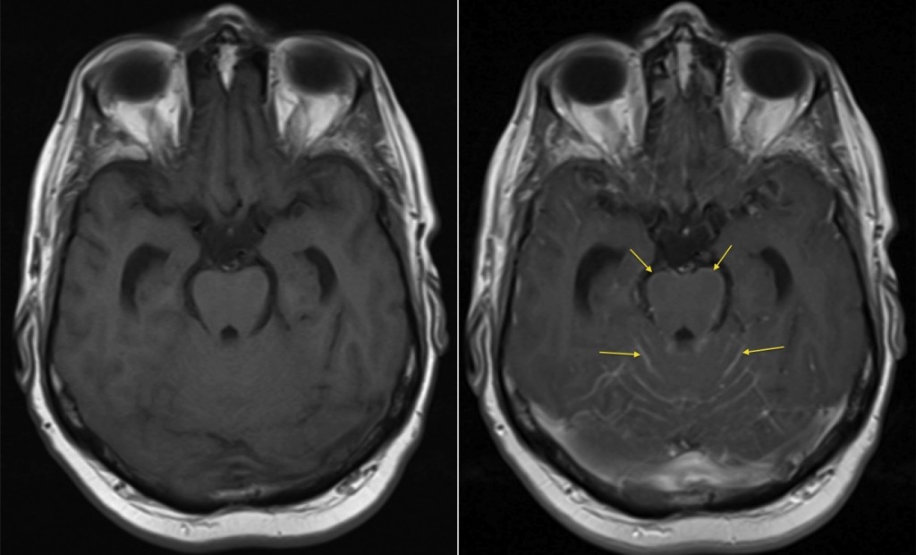
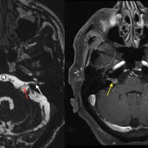
Leptomeningeal metastases. When assessing an MRI brain for metastatic disease, in addition to searching for focal lesions in the cerebrum and cerebellum, we have to look carefully at the post-contrast sequences for abnormal enhancement of the meninges due to metastatic infiltration. This can be very subtle, as in this example. The image on the left is non-contrast T1-weighted, and appears normal, however following gadolinium injection, the T1-weighted image on the right shows abnormal enhancement of the meninges around the midbrain and along the cerebellar folia (arrows). This patient had a background of lung cancer, which is one of the most common sources of meningeal metastatic disease, in addition to breast cancer and melanoma.