Multiple sclerosis – enhancing plaque
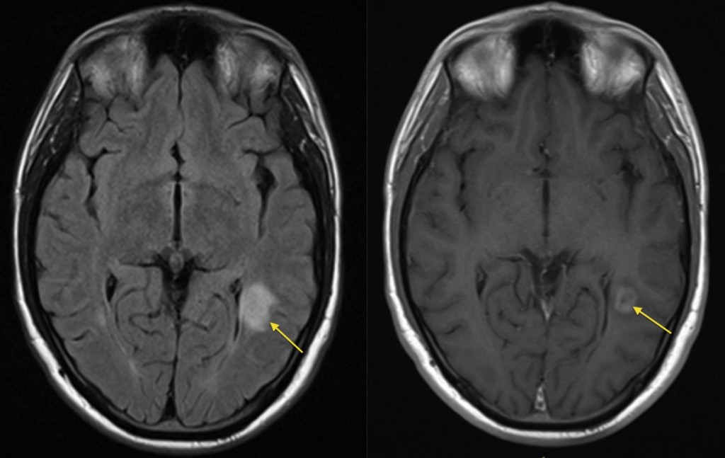
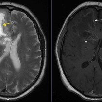
Multiple sclerosis – enhancing plaque. When evaluating an MS patient for active disease, we often perform post-contrast MR imaging. This example shows a large demyelination plaque in the left parietal lobe (arrow) on the FLAIR image on the left. On the right is a T1-weighted image obtained post gadolinium injection and shows extensive enhancement of the plaque, which tells us that the plaque is active and may therefore help guide treatment. In patients where the diagnosis of MS is uncertain, the presence of both enhancing and non-enhancing plaques helps confirm ‘dissemination in time’, one of the criteria for diagnosis.