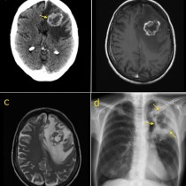
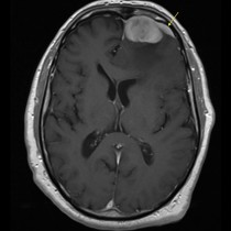
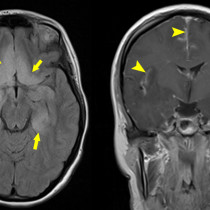
Glioblastoma multiforme
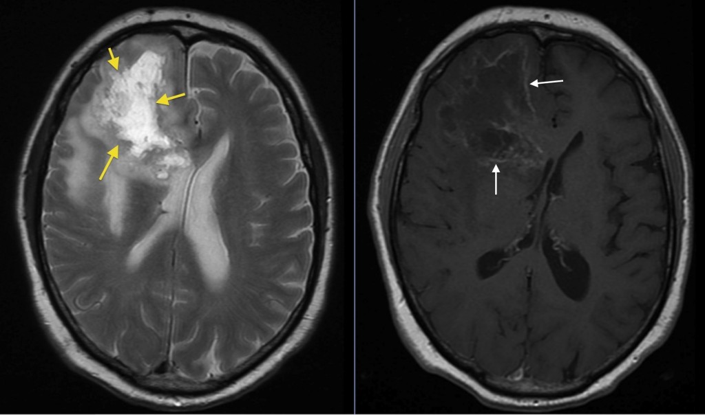
Glioblastoma multiforme. This patient presented with left sided weakness, of gradual onset. T2-weighted MRI, left, shows a large cystic mass in the right frontal lobe (arrows), surrounded by white marrow oedema (‘vasogenic oedema’, which spares the grey matter). There is some compression of the right lateral ventricle and some shift of midline structures to the left. Post-contrast T1-weighted image, right, shows peripheral enhancement of the tumour (white arrows). Biopsy confirmed glioblastoma multiforme. Although peripheral enhancement is common in cerebral metastases, the marked irregularity of the above mass would be atypical for metastatic disease.