Obstructive hydrocephalus
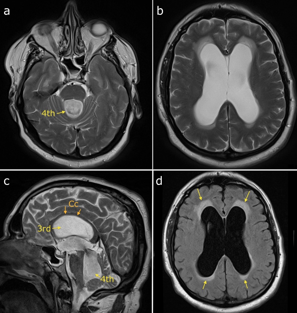
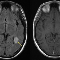
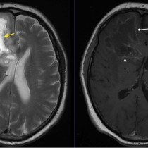
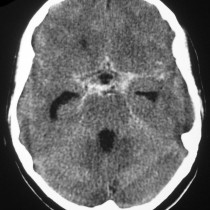
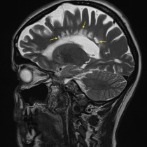
This patient presented with progressive confusion and unsteadiness. She had a background of previous TB meningitis. Transverse T2-weighted images (a) and (b) show severe dilatation of the fourth and lateral ventricles. The sagittal T2-weighted image (c) shows a grossly distended third ventricle, with elevation and thinning of the corpus callosum above it (‘Cc’). FLAIR image (d) shows abnormal high signal around the lateral ventricles, representing transependymal oedema (arrows). As all four ventricles are dilated in this case, it tells us that the site of obstruction is either in the foramina of Luschka or Magendie or in the subarachnoid space (rather than the aqueduct of Sylvius, between the third and fourth ventricles). One of the potential complications of meningitis is hydrocephalus as in this example; this can occur following viral, bacterial, mycobacterial or even sarcoid meningitis. Treatment is generally with a VP shunt.