Herpes simplex encephalitis

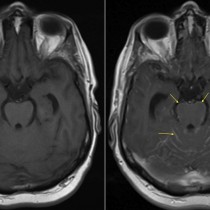
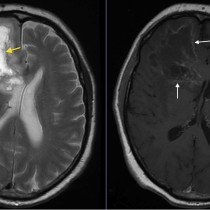
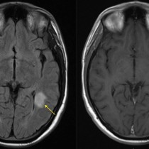
Herpes simplex encephalitis. This 26 year old woman presented with seizures. FLAIR image from her MRI, left, shows extensive abnormal high signal in the classic distribution seen in HSV encephalitis (arrows) – the medial aspects of the temporal lobes, and the inferomedial aspects of the frontal lobes. The coronal post-gadolinium T1-weighted image on the right shows abnormal meningeal thickening and enhancement, arrowheads.