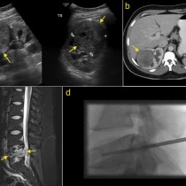
Cauda equina compression
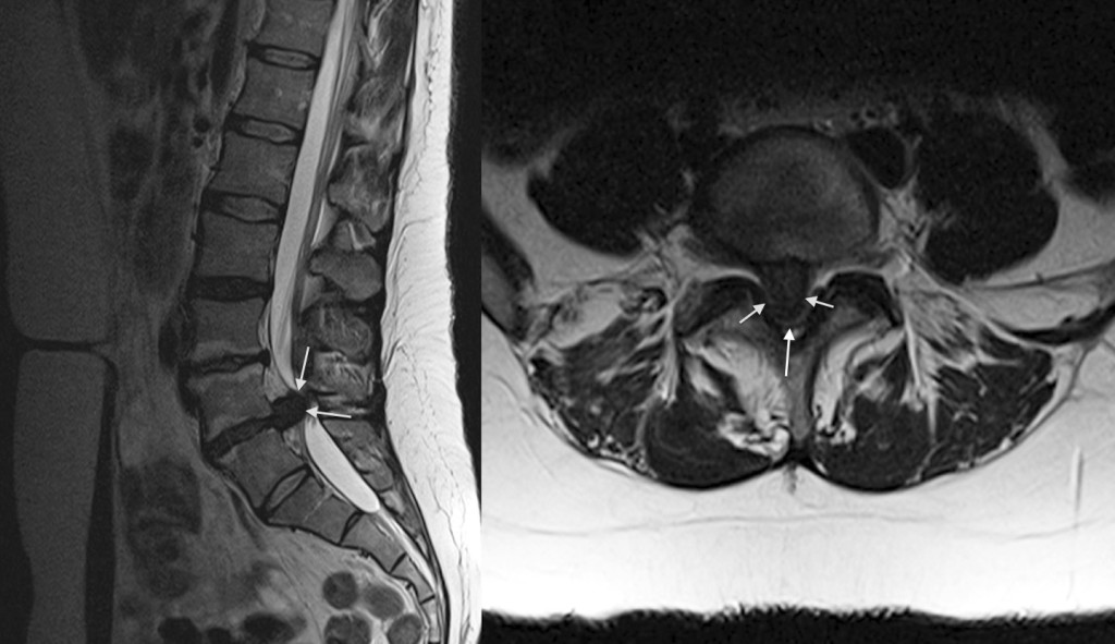
One of the most common reasons we are asked to perform emergency MRI studies is for patients who present with clinical features suggesting spinal cord or caudal equina compression. This patient arrived at the ED with sudden onset severe low back pain, saddle anaesthesia, leg weakness and urinary incontinence. Sagittal T2-weighted MRI, left, shows a large extruded low signal disk at L4-5 (arrows), with no residual CSF in the spinal canal at this level. The transverse T2-weighted image, right, shows how the disk (arrows) has completely obliterated the spinal canal – normally this image would show the roots of the cauda equina surrounded by high signal CSF. The patient was transferred to the spinal unit at the Mater Hospital for urgent decompression.