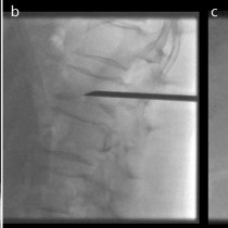
Lumbar transforaminal nerve block
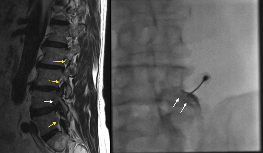
Lumbar transforaminal nerve block. This patient presented with severe left leg pain in an L4 distribution. The sagittal T2-weighted image from his MRI lumbar spine, left, shows normal appearance of most of the exiting nerve roots (yellow arrows), with the nerves surrounded by high signal fat. At L4-5 however, there is no residual high signal fat around the exiting L4 nerve root (white arrow) as it is being compressed by a combination of a disk bulge and hypertrophy of the adjacent facet joint. A nerve block was requested. The image on the right shows this being performed; the patient is placed prone, a 22-gauge spinal needle is advanced to the neural exit foramen and some iodinated contrast has been injected, outlining the nerve root (white arrows). At this point, a corticosteroid is injected along with local anaesthetic. This usually results in significant symptomatic improvement for these patients within a matter of days.