Vertebroplasty
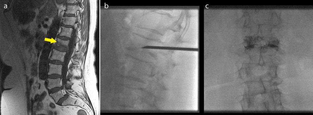
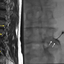
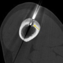
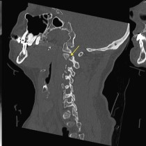
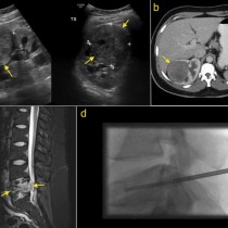
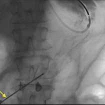
Vertebroplasty. The sagittal image from this osteoporotic patient’s MRI spine, left, shows a fracture of L1 (arrow). The patient’s pain did not respond to conservative treatment therefore vertebroplasty was arranged. In this procedure, performed under sedation, large bore (11-13G) needles are advanced through the pedicles of the affected vertebra(e) into the anterior aspect of the vertebral body, and a small quantity (usually no more than 3-4 mls) of cement injected. The lateral fluoroscopic image shows the needle in position, while the AP image on the right shows the final result, with cement filling the vertebral body. Many patients, including this one, report immediate improvement in their pain after this procedure.