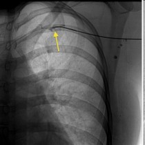
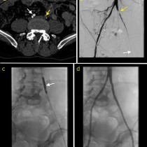
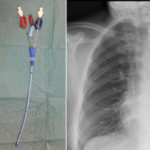
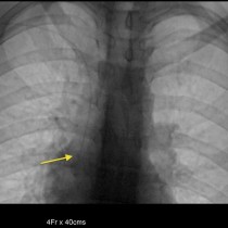
Venous thrombolysis
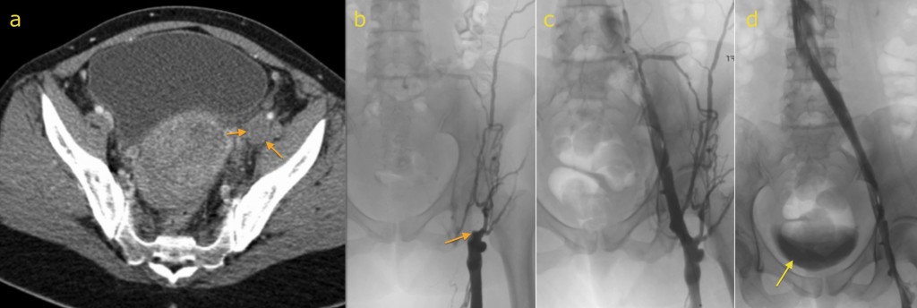
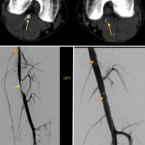
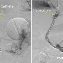
Venous thrombolysis. This young woman, recently post-partum, developed an acutely swollen left lower limb due to an ileofemoral DVT. The cause in this case was a condition called May-Thurner syndrome, in which the left common iliac vein is compressed against the lumbar spine by the right common iliac artery as it passes over it, predisposing patients to thrombosis. CT venogram (a) shows expansion of the left external iliac vein by the low density thrombus (arrows). A catheter was inserted through the left femoral vein below the thrombus, a venogram performed (showing occlusion in image (b), arrow), and thrombolysis commenced. The next day, a venogram (c) showed recanalization of the vein. In order to prevent recurrence, a metal stent was inserted into the left common iliac vein. Venogram (d) at the end of the procedure shows a widely patent left iliac vein. Note how some of the contrast injected earlier has already been excreted by the kidneys and has passed into the bladder (arrow).