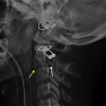
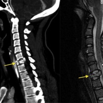
C2 fracture – XR and CT
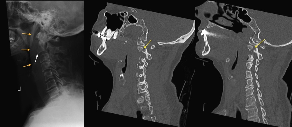
This 58 year old woman fell from a wall. Initial lateral cervical spine radiograph shows abnormal thickening of the prevertebral soft tissues anterior to the upper cervical spine (remember, from C1 to C4 the thickness of the prevertebral soft tissues should be no more than 50% of the AP dimension of the adjacent vertebral body). The soft tissue thickening is indicated by the orange arrows, and should always heighten your suspicion for an underlying fracture in a trauma setting. You will also notice that the anterior margin of the C2 vertebral body does not align normally with the body of C3 below (white arrow). The underlying fracture is difficult to spot on the radiograph but is indicated by the yellow arrow – the injury is at the junction of the pedicles and the vertebral body. This is more clearly demonstrated on the two CT images provided – both C2 pedicles had fractured, making this is a form of ‘hangman’ fracture.