• Close
    • Home
    • Videos
    • Tutorials
    • Gallery
    • Links
    • FAQs
    • All Case Studies
    • Diagnostic Imaging
      • Imaging Modalities
        • Radiography
        • Fluoroscopy
        • Ultrasound
        • CT
        • MRI
        • Nuclear Medicine
      • Contrast Media
      • Radiological Anatomy
      • Case Studies
      • PACS
      • Radiation Safety
      • Radiological Descriptive Terms
    • Interventional Radiology
      • Introduction to IR
      • Image-guided Biopsy
      • Case Studies
    • UCD Students
      • Weekly Rotation
      • Radiology Conferences
      • Resources
      • Undergraduate Medals
    • Electives
    • Careers
    • About Us
St. Vincent's University Hospital Radiology Department
  • Diagnostic Imaging
    • Imaging Modalities
      • Radiography
      • Fluoroscopy
      • Ultrasound
      • CT
      • MRI
      • Nuclear Medicine
    • Contrast Media
    • Radiological Anatomy
    • Case Studies
    • PACS
    • Radiation Safety
    • Radiological Descriptive Terms
  • Interventional Radiology
    • Introduction to IR
    • Image-guided Biopsy
    • Case Studies
  • UCD Students
    • Weekly Rotation
    • Radiology Conferences
    • Resources
    • Undergraduate Medals
  • Electives
  • Careers
  • About Us
  • Home
  • Videos
  • Tutorials
  • Gallery
  • Links
  • FAQs
  • All Case Studies

Thoracic spine fracture

Thoracic spine fracture

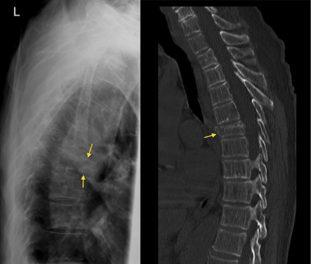
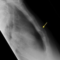
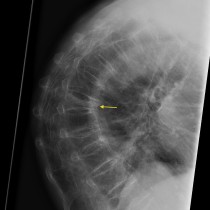
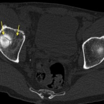
Thoracic vertebral fracture. The radiograph on the left shows some relative loss of height in the anterior aspect of T7 (arrows), consistent with a mild compression fracture. These are much easier to identify on CT (right), especially in osteoporotic patients.

Related Case Studies

  • Sternal fracture
  • Buckle fracture
  • Osteoporotic fractures
  • Acetabular fracture
  • Femoral neck fracture
  • Tripod fracture
Back
  • Interventional Radiology
  • Diagnostic Imaging
    • Emergencies
    • Neuro
    • Tubes & Lines
    • Trauma
      • Acromion Fracture
      • Scapular fracture
      • Renal laceration – CT
      • Jefferson fracture
      • Sternal fracture
      • Calcaneal Fracture
      • Posterior shoulder dislocation
      • Liposerohaemarthrosis
      • Shoulder dislocation with Bankart fracture
      • Compound patellar fracture
      • Mandibular fractures – OPG
      • Scaphoid AVN – CT
      • Penetrating chest trauma
      • Shotgun pellets
      • Stress fracture of femoral neck
      • Skull fracture – CT
      • Pneumothorax and rib fractures – CXR and CT
      • Clay shoveler’s fracture
      • Rib Fractures
      • C2 fracture – XR and CT
      • Subtalar dislocation
      • Olecranon fracture
      • Retroperitoneal haemorrhage – PFA/CT
      • Rectus sheath haematoma – CT
      • Tripod fracture
      • Femur fracture
      • Osteoporotic fractures
      • Mallet finger
      • Penetrating trauma
      • Acetabular fracture
      • Acromioclavicular separation
      • Orbital blowout fracture
      • Tripod fracture
      • Foreign bodies
      • Buckle fracture
      • Thoracic spine fracture
      • Bennett’s fracture
      • Elbow dislocation
      • Sternal fracture
      • Intertrochanteric fracture
      • Patellar fracture
      • Boxer’s fracture
      • Cervical burst fracture
      • Inferior shoulder dislocation
      • Stress fracture of tibia
      • Meniscal tear
      • ACL tear
      • Frostbite
      • Lunate dislocation
      • Periprosthetic femur fracture
      • Hangman fracture
      • Galleazzi fracture/dislocation
      • Posterior knee dislocation
      • Hip dislocation, central
      • Dislocated hip prosthesis
      • Smith’s fracture
      • Splenic laceration
      • Liver laceration
      • Pneumothorax and subcutaneous emphysema – CT
      • Shotgun injury – CT
      • Lumbar spine dislocation
      • Nail gun injury
      • Colles fracture
      • Traumatic amputation
      • Gunshot injury
      • Diaphragmatic hernia
      • Maisonneuve fracture
      • Finger dislocation
    • Oncology
    • Hepatobiliary
    • Genitourinary
    • Gastrointestinal
    • Chest
    • Musculoskeletal
    • Vascular

All Case Studies

  • Main Categories

    • Interventional Radiology
      • Vascular IR
      • Non-Vascular IR
      • IR Oncology
      • Musculoskeletal IR
    • Diagnostic Imaging
      • Emergencies
      • Neuro
      • Tubes & Lines
      • Trauma
      • Oncology
      • Hepatobiliary
      • Genitourinary
      • Gastrointestinal
      • Chest
      • Musculoskeletal
      • Vascular
  • CONTACT US

    Department of Radiology
    St Vincent’s University Hospital
    Elm Park
    Dublin 4
    Send email

    Links

    • FAQs
    • Links

    SOCIAL

    © Copyright St Vincent's University Hospital Radiology Department 2025
    Web Design Dublin

    Search St. Vincent's University Hospital Radiology Department

    This website uses cookies. Accept Read More