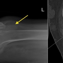
Femoral neck fracture
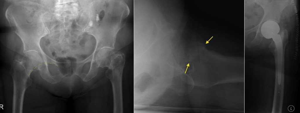
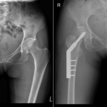
Femoral neck fracture. The AP pelvic radiograph shows a mildly displaced fracture through the base of the left femoral neck. These can be difficult to identify, however if you remember to check the integrity of Shenton’s line (illustrated on the right hip) it will help you to pick them up. The fracture line is better demonstrated on the lateral view (middle image, arrows). A significant percentage of hip fractures are radiographically-occult, however, therefore we have low threshold for performing a pelvic CT when there is clinical suspicion of a fracture but radiographs are normal. This will often demonstrate a femoral neck fracture, or an alternative fracture in the pubic rami or sacral ala (these can be very difficult to spot on plain films). In a small proportion of cases, fractures that are occult on radiographs will also be occult on CT (especially in osteoporotic patients) – in this situation, we can either perform an MRI (which will show marrow oedema at the site of the fracture), or a bone scan. The right hand image was performed later the same day, and shows that the patient has had a hemiarthroplasty.