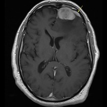
Subacute subdural haematoma – MRI
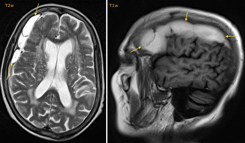
Intracranial haemorrhage undergoes a steady and predictable change in appearance on both CT and MRI. On CT, blood becomes progressively less dense and in chronic subdural haematomas can be of similar density to CSF. On MRI, the intensity of the blood changes on both T1- and T2-weighted imaging. In the acute phase (1-2 days), haemorrhage is of low to intermediate signal on both T1w and T2w, while in the subacute phase the signal on both T1w and T2w progressively increases. In this example, the haematoma is hyperintense on the left-hand T2-weighted image (arrows), and is also hyperintense on the right-hand sagittal T1-weighted image.