Venous sinus thrombosis
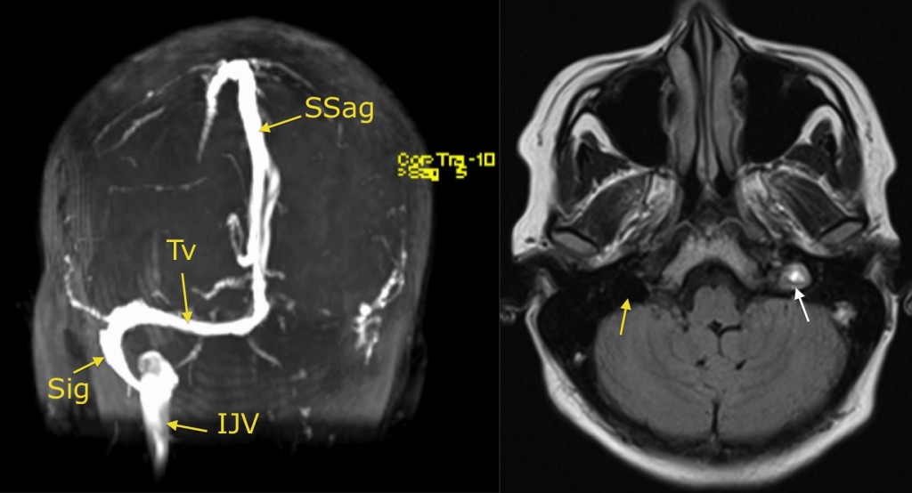
Venous sinus thrombosis. This young woman presented to the ED with severe headache. CT brain was normal, however as she was on the OCP (a risk factor for cerebral venous thrombosis) an MRI brain including an MR venogram was requested. The image on the left is a MIP (maximum intensity projection) from the MRV. You can see normal flow through the superior sagittal sinus (Sag), right transverse sinus (Tv), right sigmoid sinus (Sig) and right internal jugular vein (IJV). Normally, we should see a fairly symmetric picture on the left, however in this case no flow is evident through the left transverse or sigmoid sinuses, or in the left internal jugular. FLAIR image, right, shows normal ‘flow void’ (due to moving blood) in the right internal jugular vein (yellow arrow), but abnormal high signal in the left IJV, due to thrombus (white arrow). In addition to the OCP, risk factors for venous sinus thrombosis include pregnancy and prothrombotic conditions such as factor V Leiden. As well as MRV, the diagnosis can be made with CT Venography.