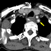
Deep venous thrombosis
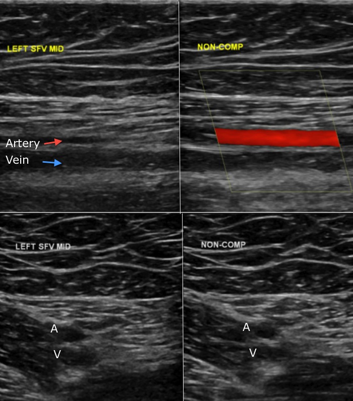
DVT. This ultrasound study shows the typical appearances of a DVT, in this case in the superficial femoral vein. While we can use colour Doppler to illustrate whether or not there is flowing blood in the vein, the most accurate method of confirming or excluding a DVT is to use the ultrasound probe to put pressure on the vein – in the normal setting the vein will collapse fully, however if there is thrombus in it then it will not be possible to fully compress it. The top images in this example show a longitudinal view of the femoral vein, with compression used on the right-hand image, while the bottom row shows transverse images, again with compression used on the right.