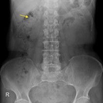
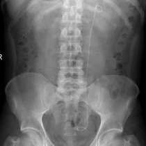
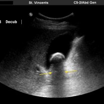
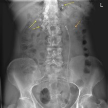
Ureteric calculus on CT KUB
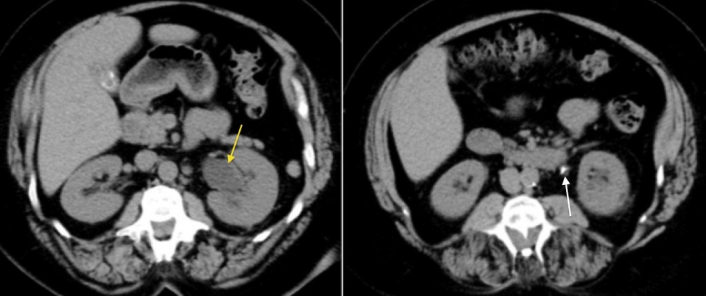
Ureteric calculus. These images from a CT KUB (kidneys, ureters, bladder – performed without IV contrast) show left hydronephrosis (yellow arrow), caused by a calculus in the proximal ureter (white arrow). CT KUB is a rapid way of establishing the presence or absence of renal and ureteric calculi, and will also show alternative causes of symptoms when no calculi are identified.