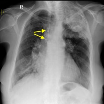
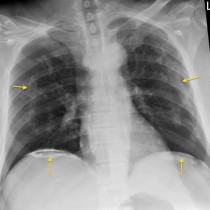
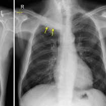
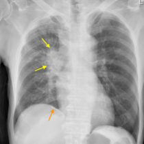
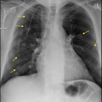
Thoracoplasty – chest radiograph
This bizarre appearance is something we don’t see very often these days, but was for a long time a very popular treatment option for patients with advanced tuberculosis. This patient is 86 years old, and the chest radiograph was performed for suspected pneumonia (no consolidation is present here though).
Thoracoplasty involved the removal of several ribs from the worst-affected side of the chest, with the goal being to cause the affected portion of the lung to collapse, arresting the progression of disease.
In the procedure, resection of multiple ribs was performed, beginning at the second rib and working down as far as was necessary to cause collapse of the desired part of the lung. Usually this meant five or six ribs being removed, however in some patients additional ribs were resected below this level. Somewhat unsettlingly, the procedure was often performed under local anaesthetic in the early 20th century.
The radiographic result of this procedure is characteristic: the chest wall is grossly deformed, the upper lobe of the lung is collapsed and the overlying pleura becomes densely calcified.
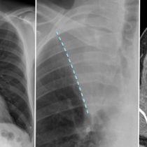
Another obsolete method for treating tuberculosis, with the same desired outcome of collapsing the affected lung, was to place plastic balls outside the pleura. See an example of this here.
While these procedures are no longer performed, we occasionally see elderly patients coming through the department for imaging who have had a thoracoplasty in their youth.
Reference: Whittemore W. Thoracoplasty in pulmonary tuberculosis. NEJM 1932;207:211-217.