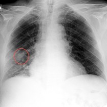
Lung cancer in patient with previous asbestos exposure
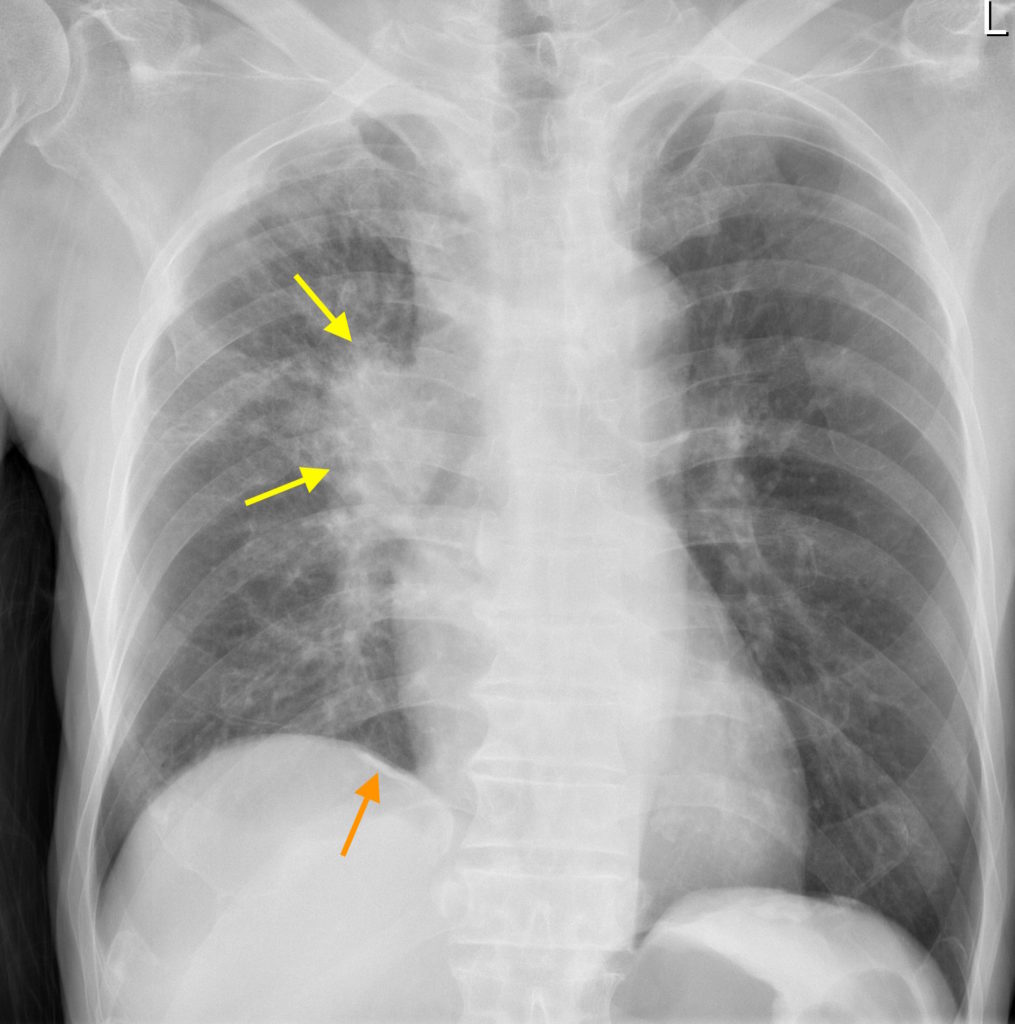
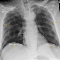
This male patient, a smoker, presented with haemoptysis. Chest X-ray shows a large mass at the right hilum (yellow arrows). You will also notice that there is a calcified pleural plaque adjacent to the medial aspect of the right hemidiaphragm (orange arrow), indicating previous asbestos exposure. Biopsy of the hilar mass showed non-small cell lung cancer.
While the patient’s history of smoking would, on its own, be sufficient to induce a lung neoplasm, as you are aware prior asbestos exposure has a synergistic effect in smokers and further increases the risk of developing lung cancer.
Potential imaging findings in the chest in patients with a history of asbestos exposure include:
- Pleural plaques
- Diffuse pleural thickening
- Mesothelioma
- Interstitial lung disease (‘asbestosis’, predominantly in the lung bases)
- Rounded atelectasis (focal fibrosis and volume loss in a lung, often mass-like)
- Bronchogenic carcinoma