Pancoast tumour
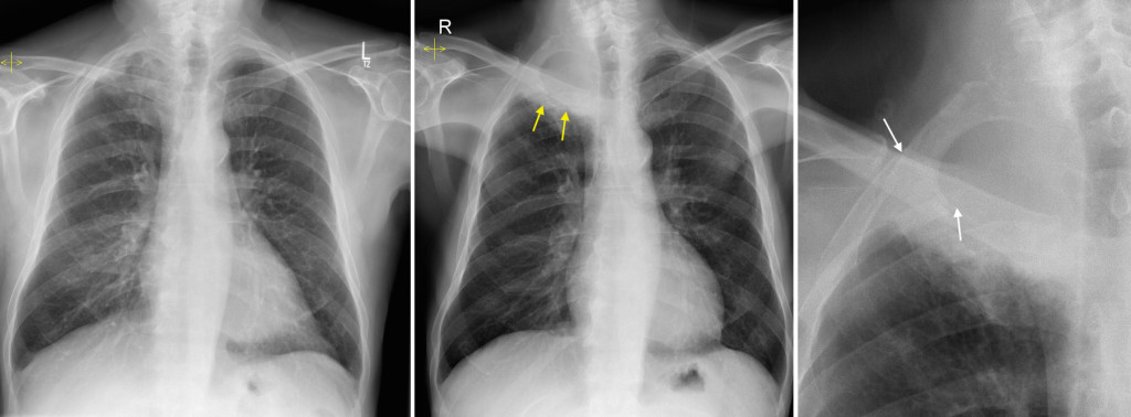
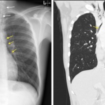
This elderly man presented to the ED with a cough and was also complaining of right shoulder pain. His CXR (middle image) shows a large right apical mass (yellow arrows) that was not present on a previous study from 10 months earlier (left image). On the magnified image on the right, you can see that the mass has eroded the posterior aspects of the second and third ribs (the ribs disappear abruptly at the sites of the white arrows).
Pancoast tumours are usually non-small cell lung cancers and may present with chest, shoulder and/or arm pain, as well as (classically, but less than 50% of the time in reality) Horner syndrome. Although CT is routinely the next imaging step when one of these lesions is identified on a chest radiograph, assessing the presence of involvement of the brachial plexus (and, therefore, resectability of the tumour) is best performed with MRI.