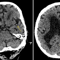
Haemorrhagic transformation of CVA
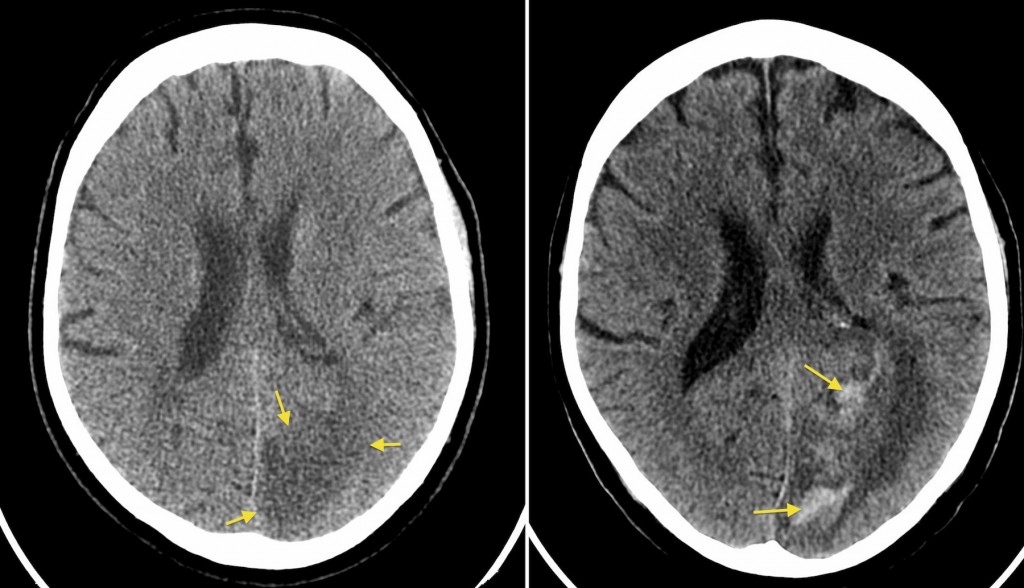
The image on the left is from this patient’s CT performed following presentation to the ED. It shows a large area of low attenuation with loss of grey-white matter differentiation in the left occipital lobe, due to an infarct. The patient deteriorated three days later and a repeat CT (right) showed haemorrhagic transformation, with extensive high attenuation haemorrhage within the infarct (arrows). Note the increased mass effect on the left lateral ventricle. Haemorrhagic transformation tends to occur in the first few days after an infarct, and is of course more common in patients who are anticoagulated or treated with thrombolysis.