• Close
    • Home
    • Videos
    • Tutorials
    • Gallery
    • Links
    • FAQs
    • All Case Studies
    • Diagnostic Imaging
      • Imaging Modalities
        • Radiography
        • Fluoroscopy
        • Ultrasound
        • CT
        • MRI
        • Nuclear Medicine
      • Contrast Media
      • Radiological Anatomy
      • Case Studies
      • PACS
      • Radiation Safety
      • Radiological Descriptive Terms
    • Interventional Radiology
      • Introduction to IR
      • Image-guided Biopsy
      • Case Studies
    • UCD Students
      • Weekly Rotation
      • Radiology Conferences
      • Resources
      • Undergraduate Medals
    • Electives
    • Careers
    • About Us
St. Vincent's University Hospital Radiology Department
  • Diagnostic Imaging
    • Imaging Modalities
      • Radiography
      • Fluoroscopy
      • Ultrasound
      • CT
      • MRI
      • Nuclear Medicine
    • Contrast Media
    • Radiological Anatomy
    • Case Studies
    • PACS
    • Radiation Safety
    • Radiological Descriptive Terms
  • Interventional Radiology
    • Introduction to IR
    • Image-guided Biopsy
    • Case Studies
  • UCD Students
    • Weekly Rotation
    • Radiology Conferences
    • Resources
    • Undergraduate Medals
  • Electives
  • Careers
  • About Us
  • Home
  • Videos
  • Tutorials
  • Gallery
  • Links
  • FAQs
  • All Case Studies

Acute infarct

Acute infarct

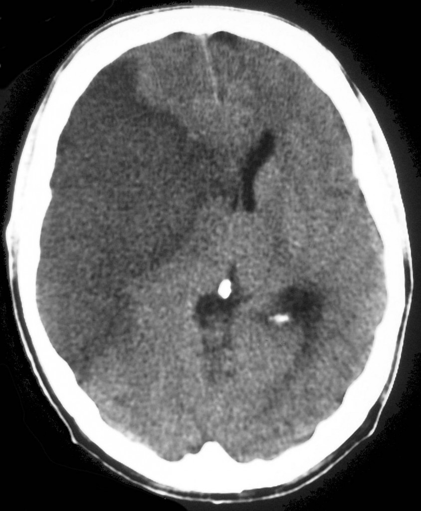
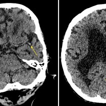
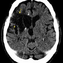
Acute infarct. This noncontrast CT image shows a large area of abnormal low attenuation in the territory of the right middle cerebral artery, representing an acute infarct. Remember, however, that CT is usually normal in the first few hours after a stroke.

Related Case Studies

  • Acute infarct – CT
  • Acute MCA infarct – dot sign
  • Cerebellar infarct
  • Haemorrhagic transformation of CVA
  • Acute infarct with dense MCA sign
  • Old CVA
Back
  • Interventional Radiology
  • Diagnostic Imaging
    • Emergencies
    • Neuro
      • Persistent metopic suture
      • Acute on chronic subdural haematoma
      • Cerebral Amyloid Angiopathy – MRI
      • Ruptured pericallosal aneurysm with subarachnoid haemorrhage – CT
      • Cervical myelopathy – MRI
      • Occluded middle cerebral artery on CT angiography
      • Lumbar spinal stenosis
      • Encephalomalacia due to old MCA infarct
      • Hypertensive intracerebral haemorrhage
      • Chiari I malformation with syrinx
      • Mesial temporal sclerosis
      • Ocular melanoma – US and MRI
      • Obstructive hydrocephalus
      • Subacute subdural haematoma – MRI
      • Haemorrhagic transformation of CVA
      • Acute MCA infarct – dot sign
      • Global hypoxic ischaemic injury
      • Herpes simplex encephalitis
      • Syrinx – MRI
      • Huge subarachnoid haemorrhage – CT angio
      • Acute infarct – CT
      • Thyroid eye disease
      • Subacute combined degeneration of the cord
      • Normal pressure hydrocephalus
      • Meningioma
      • Meningeal metastases
      • Benign intracranial hypertension
      • Glioblastoma multiforme
      • Colloid cyst
      • Acoustic neuroma
      • Venous sinus thrombosis
      • Dural AVM
      • Embolic infarcts
      • Cerebral contusion
      • Cerebellar infarct
      • Intracerebral haemorrhage
      • Multiple sclerosis – enhancing plaque
      • Multiple sclerosis – spinal cord atrophy
      • Old CVA
      • Brain metastasis
      • Chronic subdural haematoma
      • Multiple sclerosis
      • Acute subdural haematoma
      • Acute infarct with dense MCA sign
      • Berry aneurysm
      • Acute subarachnoid haemorrhage (2)
      • Acute subarachnoid haemorrhage
      • Acute infarct
      • Extradural haematoma
    • Tubes & Lines
    • Trauma
    • Oncology
    • Hepatobiliary
    • Genitourinary
    • Gastrointestinal
    • Chest
    • Musculoskeletal
    • Vascular

All Case Studies

  • Main Categories

    • Interventional Radiology
      • Vascular IR
      • Non-Vascular IR
      • IR Oncology
      • Musculoskeletal IR
    • Diagnostic Imaging
      • Emergencies
      • Neuro
      • Tubes & Lines
      • Trauma
      • Oncology
      • Hepatobiliary
      • Genitourinary
      • Gastrointestinal
      • Chest
      • Musculoskeletal
      • Vascular
  • CONTACT US

    Department of Radiology
    St Vincent’s University Hospital
    Elm Park
    Dublin 4
    Send email

    Links

    • FAQs
    • Links

    SOCIAL

    © Copyright St Vincent's University Hospital Radiology Department 2023
    Web Design Dublin

    Search St. Vincent's University Hospital Radiology Department

    This website uses cookies. Accept Read More