Old CVA
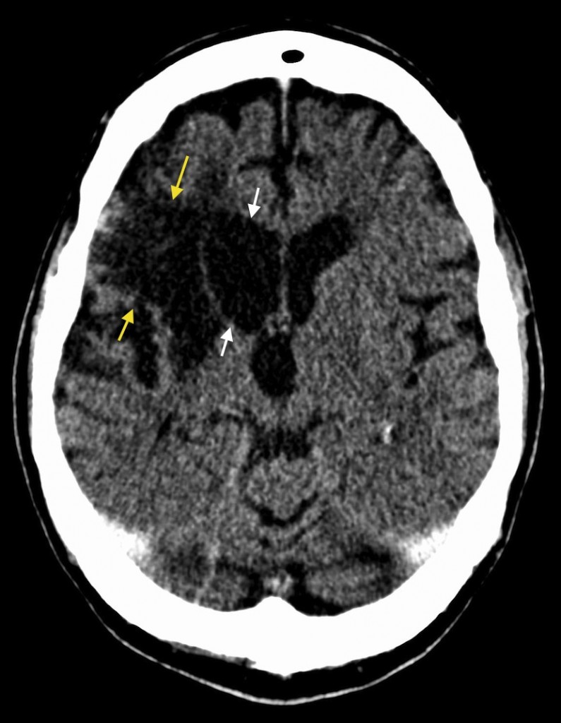
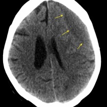
Old CVA. Acute infarcts can be difficult to identify on CT as the degree of density reduction in the infarcted tissue can be minimal. In contrast, in an old CVA the infarcted area becomes of similar density to CSF, as in this case of a longstanding right MCA infarct (yellow arrows). In addition, we also see evidence of loss of tissue volume – the opposite of mass effect – which results in widening of the sulci and enlargement of the nearby ventricle (white arrows). This is called ‘ex-vacuo dilatation’.