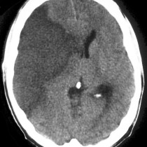
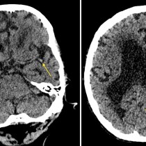
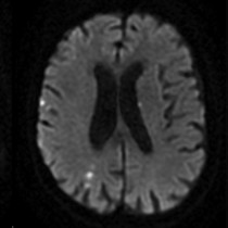
Cerebellar infarct
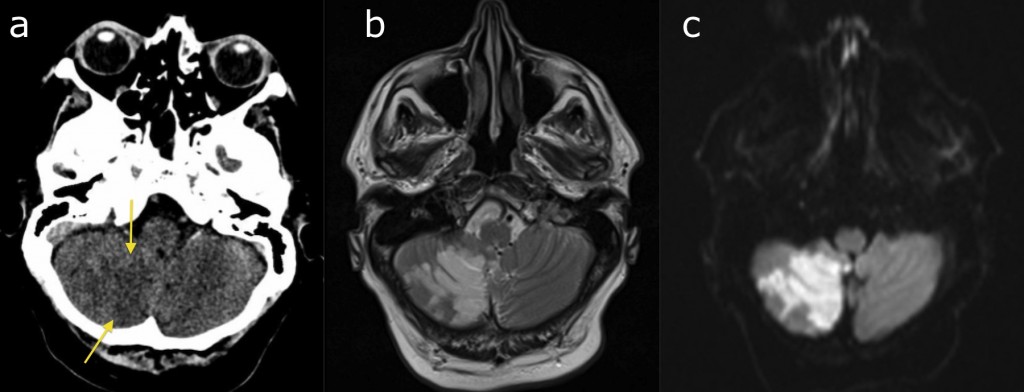
Cerebellar infarct. It can be difficult to assess the inferior aspect of the cerebellum on CT due the dense bone in the surrounding skull base. In this example, there is very subtle low attenuation in the right cerebellar hemisphere (arrows), but this would be easy to overlook. The patient had clear signs of a cerebellar infarct however, so an MRI was requested – the abnormality is much more obvious on the middle T2w image, while the right-hand diffusion-weighted image shows that there is restricted diffusion at the site, confirming that it is an acute infarct.