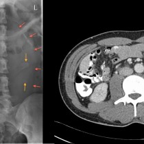
Rectus sheath haematoma – CT
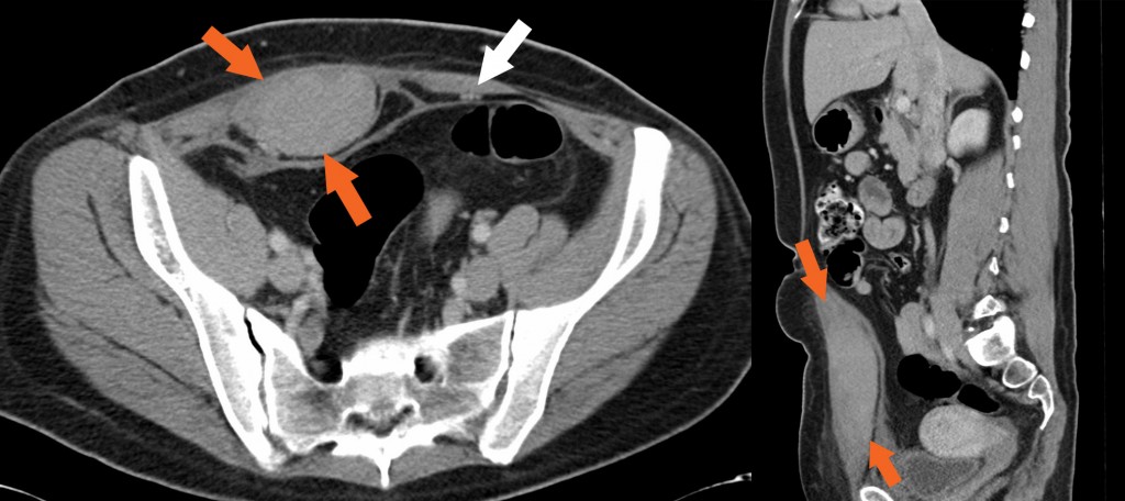
Rectus sheath haematoma. This 42 year old man was working out in a gym when he ‘pulled a muscle’ in his abdomen. He developed severe pain and swelling and presented to the ED. CT shows that the right rectus abdominus muscle is markedly enlarged (orange arrows) compared to the normal left (white arrow), and is also of much higher attenuation. This is due to a haematoma, the full length of which is illustrated on the sagittal image on the right. While in this case the haemorrhage occurred due to trauma, the rectus sheath is also a very common location for spontaneous haemorrhage in patients who are anticoagulated. Such patients can also develop spontaneous haemorrhage into the psoas muscles, and can lose significant (potentially fatal) quantities of blood as a result. In severe cases, ongoing haemorrhage into the rectus or psoas can be treated with embolization in Interventional Radiology.