Dermatomyositis – MRI
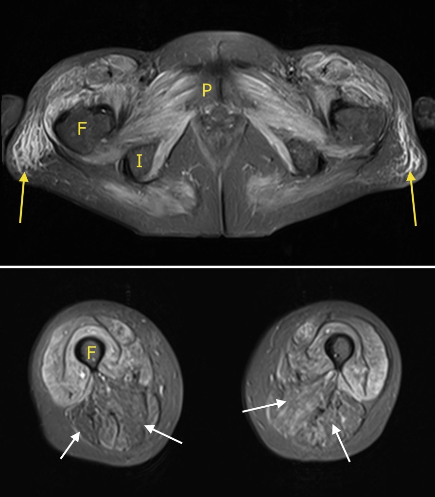
These are transverse T2-weighted fat-saturated (oedema-sensitive) MR images from a 65 year old woman who presented with severe lower limb weakness and an elevated creatine kinase. The top image is just below the hips, the lower image is at the mid-thigh level. The MR confirmed the clinical suspicion of dermatomyositis – there is severe, extensive bilaterally symmetric oedema in the muscles of the pelvic girdle as well as in the quadriceps muscles. There is also subcutaneous oedema lateral to the hips (yellow arrows). The hamstring muscles are relatively spared (white arrows). F = femur, P = pubic bone, I = ischium.