Crohn disease – MRI
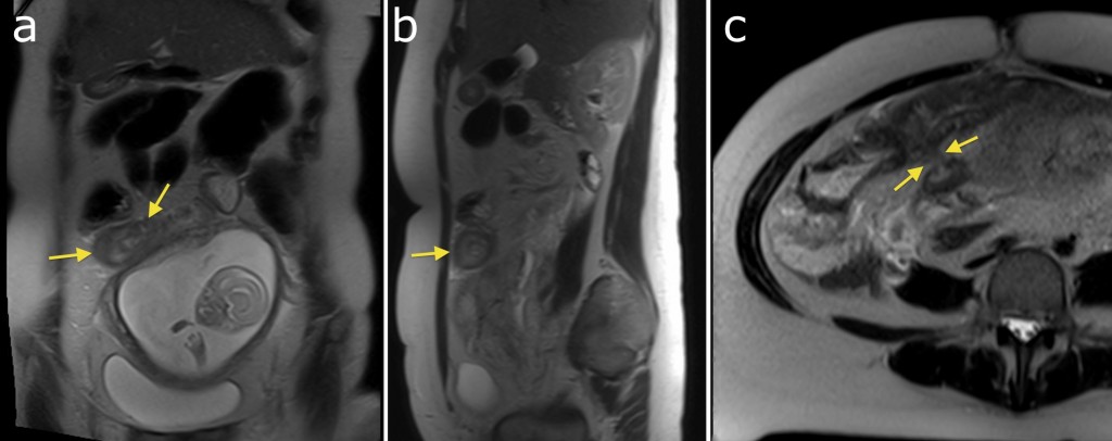
This young woman with a past history of Crohn disease presented to the ED with abdominal pain in her second trimester of pregnancy. MRI was performed – note the gravid uterus on image (a). The coronal and sagittal T2-weighted images (a) and (b) show circumferential thickening of a loop of small bowel (arrows) with some high signal oedema around them. Magnified view from a transverse image (c) showed a fistula (arrow) between two loops of small bowel. The features are those of active Crohn disease.