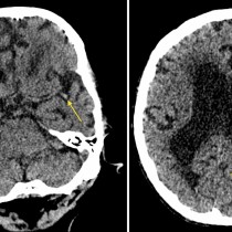
• Close
    • Home
    • Videos
    • Tutorials
    • Gallery
    • Links
    • FAQs
    • All Case Studies
    • Diagnostic Imaging
      • Imaging Modalities
        • Radiography
        • Fluoroscopy
        • Ultrasound
        • CT
        • MRI
        • Nuclear Medicine
      • Contrast Media
      • Radiological Anatomy
      • Case Studies
      • PACS
      • Radiation Safety
      • Radiological Descriptive Terms
    • Interventional Radiology
      • Introduction to IR
      • Image-guided Biopsy
      • Case Studies
    • UCD Students
      • Weekly Rotation
      • Radiology Conferences
      • Resources
      • Undergraduate Medals
    • Electives
    • Careers
    • About Us
St. Vincent's University Hospital Radiology Department
  • Diagnostic Imaging
    • Imaging Modalities
      • Radiography
      • Fluoroscopy
      • Ultrasound
      • CT
      • MRI
      • Nuclear Medicine
    • Contrast Media
    • Radiological Anatomy
    • Case Studies
    • PACS
    • Radiation Safety
    • Radiological Descriptive Terms
  • Interventional Radiology
    • Introduction to IR
    • Image-guided Biopsy
    • Case Studies
  • UCD Students
    • Weekly Rotation
    • Radiology Conferences
    • Resources
    • Undergraduate Medals
  • Electives
  • Careers
  • About Us
  • Home
  • Videos
  • Tutorials
  • Gallery
  • Links
  • FAQs
  • All Case Studies

Cerebral contusion

Cerebral contusion

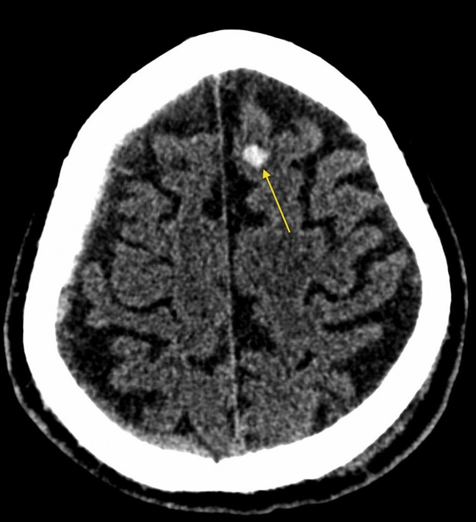
Cerebral contusion. The small intracerebral high density lesion (arrow) in this patient’s left frontal lobe on non-contrast CT occurred following a fall while intoxicated, and is typical of a contusion. These often occur in the subcortical white matter, as in this case.

Related Case Studies

  • Haemorrhagic transformation of CVA
  • Acute MCA infarct – dot sign
  • Meningioma
  • Glioblastoma multiforme
  • Acute infarct – CT
  • Global hypoxic ischaemic injury
Back
  • Interventional Radiology
  • Diagnostic Imaging
    • Emergencies
    • Neuro
      • Persistent metopic suture
      • Acute on chronic subdural haematoma
      • Cerebral Amyloid Angiopathy – MRI
      • Ruptured pericallosal aneurysm with subarachnoid haemorrhage – CT
      • Cervical myelopathy – MRI
      • Occluded middle cerebral artery on CT angiography
      • Lumbar spinal stenosis
      • Encephalomalacia due to old MCA infarct
      • Hypertensive intracerebral haemorrhage
      • Chiari I malformation with syrinx
      • Mesial temporal sclerosis
      • Ocular melanoma – US and MRI
      • Obstructive hydrocephalus
      • Subacute subdural haematoma – MRI
      • Haemorrhagic transformation of CVA
      • Acute MCA infarct – dot sign
      • Global hypoxic ischaemic injury
      • Herpes simplex encephalitis
      • Syrinx – MRI
      • Huge subarachnoid haemorrhage – CT angio
      • Acute infarct – CT
      • Thyroid eye disease
      • Subacute combined degeneration of the cord
      • Normal pressure hydrocephalus
      • Meningioma
      • Meningeal metastases
      • Benign intracranial hypertension
      • Glioblastoma multiforme
      • Colloid cyst
      • Acoustic neuroma
      • Venous sinus thrombosis
      • Dural AVM
      • Embolic infarcts
      • Cerebral contusion
      • Cerebellar infarct
      • Intracerebral haemorrhage
      • Multiple sclerosis – enhancing plaque
      • Multiple sclerosis – spinal cord atrophy
      • Old CVA
      • Brain metastasis
      • Chronic subdural haematoma
      • Multiple sclerosis
      • Acute subdural haematoma
      • Acute infarct with dense MCA sign
      • Berry aneurysm
      • Acute subarachnoid haemorrhage (2)
      • Acute subarachnoid haemorrhage
      • Acute infarct
      • Extradural haematoma
    • Tubes & Lines
    • Trauma
    • Oncology
    • Hepatobiliary
    • Genitourinary
    • Gastrointestinal
    • Chest
    • Musculoskeletal
    • Vascular

All Case Studies

  • Main Categories

    • Interventional Radiology
      • Vascular IR
      • Non-Vascular IR
      • IR Oncology
      • Musculoskeletal IR
    • Diagnostic Imaging
      • Emergencies
      • Neuro
      • Tubes & Lines
      • Trauma
      • Oncology
      • Hepatobiliary
      • Genitourinary
      • Gastrointestinal
      • Chest
      • Musculoskeletal
      • Vascular
  • CONTACT US

    Department of Radiology
    St Vincent’s University Hospital
    Elm Park
    Dublin 4
    Send email

    Links

    • FAQs
    • Links

    SOCIAL

    © Copyright St Vincent's University Hospital Radiology Department 2023
    Web Design Dublin

    Search St. Vincent's University Hospital Radiology Department

    This website uses cookies. Accept Read More