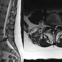
Lumbar spinal stenosis
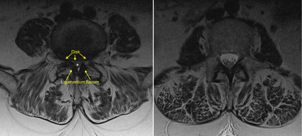
This elderly woman presented with progressive lower limb weakness on a background of chronic low back pain. She was referred for an MRI which showed severe spinal stenosis at L4-5, causing compression of the thecal sac and of the nerve roots of the cauda equina. In this transverse T2-weighted image, the L4-5 level is on the left and shows the reasons for her spinal stenosis. She has a degenerative disk bulge (‘disk’) compromising the anterior aspect of the spinal canal, while posteriorly the canal is narrowed by a combination of hypertrophied, degenerative facet joints and hypertrophied ligamenta flavum. The roots of the caudal equina (‘*’) are being compressed and there is no longer any bright CSF around them. For comparison, look at the image on the right which is from the same patient but at the L2-3 level – the nerve roots are clearly identifiable as individual structures and there is plenty of bright CSF in the spinal canal.