Percutaneous nephrostomy
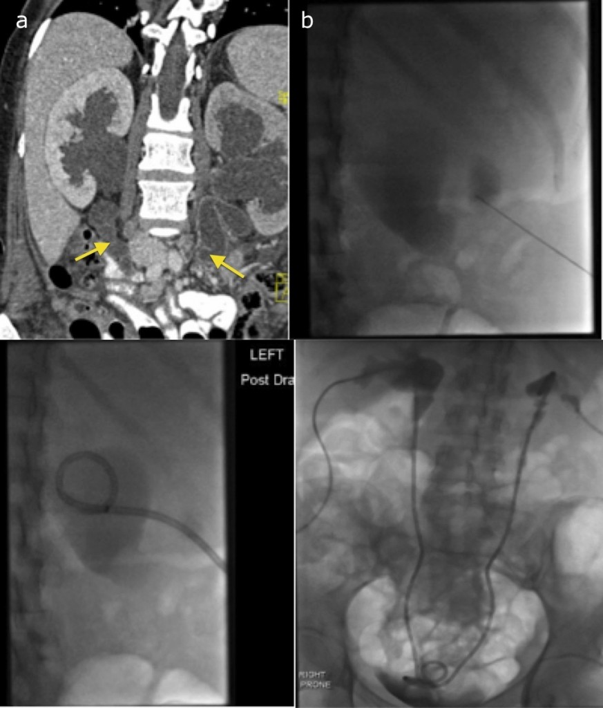
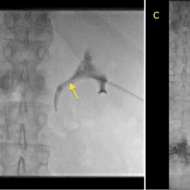
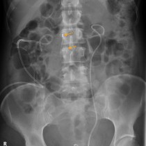
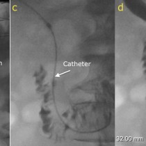
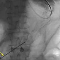
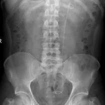
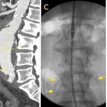
Percutaneous nephrostomy. This patient developed renal failure due to bilateral ureteric obstruction caused by inoperable pelvic recurrence of rectal cancer. The initial CT (a) shows severe bilateral hydronephrosis with dilated ureters (arrows). Using ultrasound for guidance, a needle was inserted into one of the dilated calyces in the left kidney. We then switched to fluoroscopy, and some iodinated contrast was injected through the needle to confirm that it was in the collecting system (b). A guide-wire was inserted through the needle, over which a plastic pigtail catheter was advanced into the renal pelvis (c). This was repeated on the opposite side, and urine drained from both catheters into drainage bags. The patient subsequently returned to IR for ureteric stent insertion; using the nephrostomy catheter for access, a guide wire was advanced through the ureters into the bladder, over which plastic ureteric stents were inserted (demonstrated on image (d)). 24 hours later, the nephrostomy tubes were removed. Often, percutaneous nephrostomy and ureteric stent insertion are performed at the same time (see elsewhere in this section).