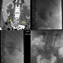
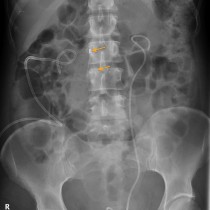
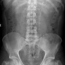
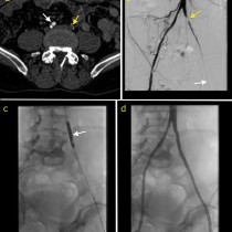
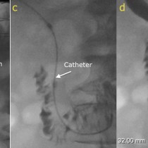
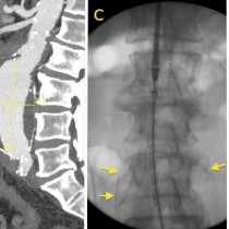
Ureteric stent insertion
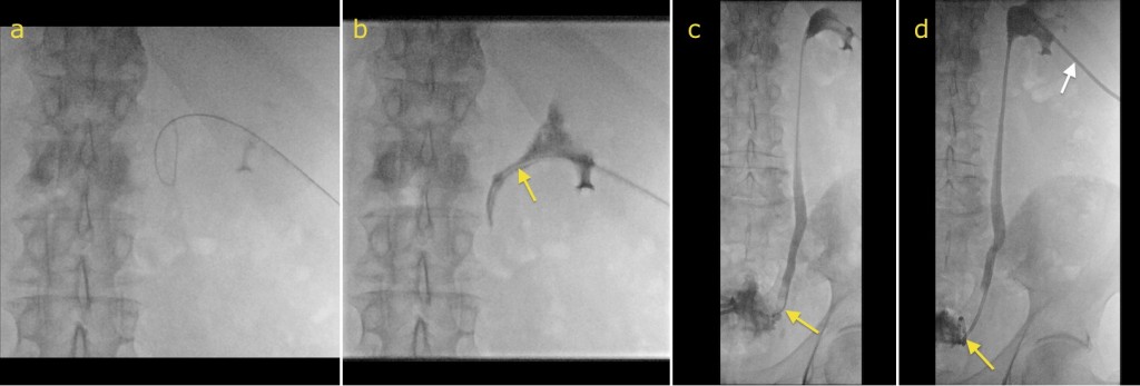
Ureteric stent insertion. In this example, the patient had bilateral hydronephrosis due to obstruction of the distal ureters by a large prostate carcinoma. Bilateral ureteric stents were requested. Image (a) shows a guidewire in the renal pelvis, which has been inserted through a needle that was guided into the kidney using visualization with ultrasound. A catheter was then inserted over the guidewire, arrow on image (b), and then advanced with a guidewire through the remainder of the ureter and into the bladder (arrow on image (c)). Once the guidewire has been safely passed across the obstruction into the bladder, a stent can be inserted over it (yellow arrow, image (d)). A nephrostomy catheter is left in the renal pelvis above the stent (white arrow), and will be removed after a day or two when we are satisfied that the stent is functioning satisfactorily.