Endovascular aneurysm repair (EVAR)
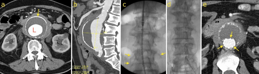
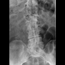
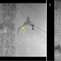
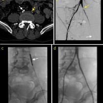
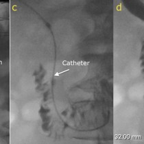
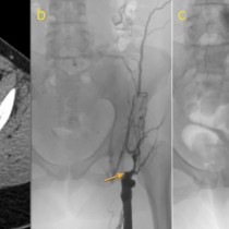
Endovascular aneurysm repair. This man presented with renal failure which was caused by ureteric obstruction, secondary to fibrosis around a huge abdominal aortic aneurysm. His ureters were stented, following which his renal function recovered, and he was then worked up for endovascular aneurysm repair. On image (a), you can see the dilated lumen of the aorta (L), surrounded by low density mural thrombus, surrounded by the calcified aortic wall (arrow) and in turn surrounded by soft tissue which represented fibrosis and which was engulfing both ureters. Image (b) is a sagittal CT reconstruction and shows the full dimensions of the aneurysm (10.9 cm in length, 7.2 cm in the anteroposterior direction). The EVAR delivery device has been inserted through the left femoral artery on image (c); you can just about make out the calcified walls of the AAA on this fluoro image (arrows). Once it is in the correct position, the covered metal stent is deployed in the aorta (d) and one or both iliac arteries. Follow-up CT (e) shows contrast flowing through the stent (arrows). Not all AAA patients are suitable for EVAR, because of the morphology of the aneurysm. However, when suitable, EVAR is an excellent alternative to open repair – the expected hospital stay post-procedure is only 2 days (compared to almost 2 weeks for open surgery), and the mortality rate is less than half of that of open aneurysm repair.