Popliteal angioplasty – atherosclerosis
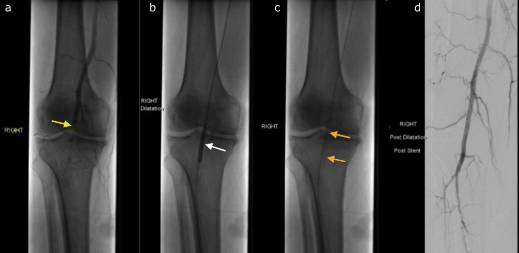
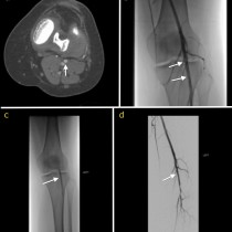
Popliteal angioplasty. This patient had symptoms of claudication in the right leg. A CT angiogram showed severe atherosclerotic stenosis of the popliteal artery, and the patient was referred for IR management. Initial angiogram (a), performed by advancing a catheter into the femoral artery, confirms the stenosis (arrow). A guide wire was passed through the stenosed segment, and an angioplasty balloon inflated (b, arrow). A metal stent was then inserted at this site (c, arrows). Subsequent angiogram (d) shows that the popliteal artery is now widely patent. This final angiographic image is from what is called a ‘digital subtraction angiogram’, or DSA – this technique subtracts the bones from the image to make it easier to see the contrast in the arteries.