Pancoast tumour – CT/MRI
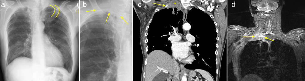
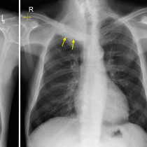
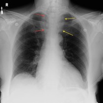
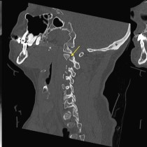
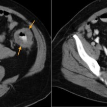
This male ex-smoker presented with new onset of right arm pain. Chest X-ray (a) shows subtle soft tissue at the right lung apex. If you look carefully, you’ll notice that the right first rib appears to have vanished – the left first rib is outlined by the yellow lines for comparison. The abnormal soft tissue is easier to see on the magnified view (b), arrow. Findings, together with the patient’s history, were suspicious for a Pancoast tumour and a CT was recommended. Also note the elevated right hemidiaphragm – phrenic nerve involvement can be a feature of Pancoast tumours. The CT (c) confirms the apical tumour (*). You can also appreciate how it is eroding adjacent rib (arrow). To better assess the relationship of such tumours to the brachial plexus, an MRI is typically performed in these patients. The tumour is indicated by the arrows on coronal STIR (fat-suppressed) MRI (d). The MRI confirmed invasion of the right brachial plexus and also showed encasement of upper thoracic nerve roots.