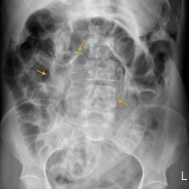
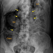
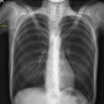
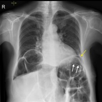
Falciform ligament sign
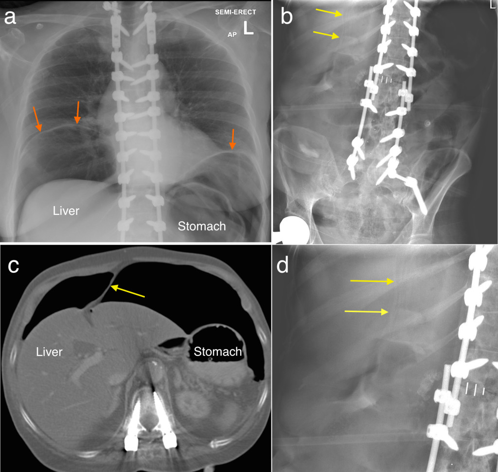
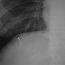
This is another sign to look out for in patients with suspected perforation – you won’t see it in isolation, as it takes a large volume pneumoperitoneum to produce the falciform ligament sign so you’ll almost certainly have already picked it up on an erect CXR or as a Rigler’s sign on a PFA. In this particular example, the erect CXR shows a huge amount of free air below both hemidiaphragms (arrows), outlining the liver and stomach (labelled on image (a)). The linear density indicated by the yellow arrows on image (b) and (magnified) on image (d) represents the falciform ligament, with air either side of it. To show you what this is caused by, a CT image from the same patient is provided (c) showing the ligament extending from the liver to the abdominal wall. The more observant among you may have noticed the extensive spinal hardware.